
Bipartite hallux sesamoid Image
Browse 85 left foot xray photos and images available, or start a new search to explore more photos and images. Browse Getty Images' premium collection of high-quality, authentic Left Foot Xray stock photos, royalty-free images, and pictures. Left Foot Xray stock photos are available in a variety of sizes and formats to fit your needs.

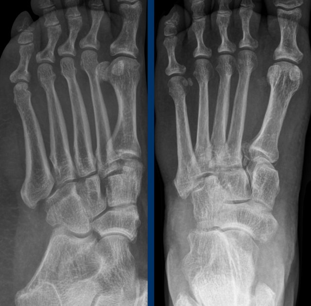
Plain radiograph (AP and lateral oblique) of the left foot (injured)... Download Scientific
Find Left Foot Xray stock images in HD and millions of other royalty-free stock photos, 3D objects, illustrations and vectors in the Shutterstock collection. Thousands of new, high-quality pictures added every day.
Normal Left Ankle Xray
A foot X-ray is a test that produces an image of the anatomy of your foot. Your healthcare provider may use foot X-rays to diagnose and treat health conditions in your foot or feet. Foot X-rays are a simple, quick, and painless process. Your leg will be positioned on an X-ray table by a radiologic technician who will then take numerous images.

Normal ankle series Image
A foot X-ray is a test that produces an image of the anatomy of your foot. Your healthcare provider may use foot X-rays to diagnose and treat health conditions in your foot or feet. Foot X-rays are quick, easy and painless procedures. A radiologic technologist will place your leg on an X-ray table and then take multiple pictures of it.
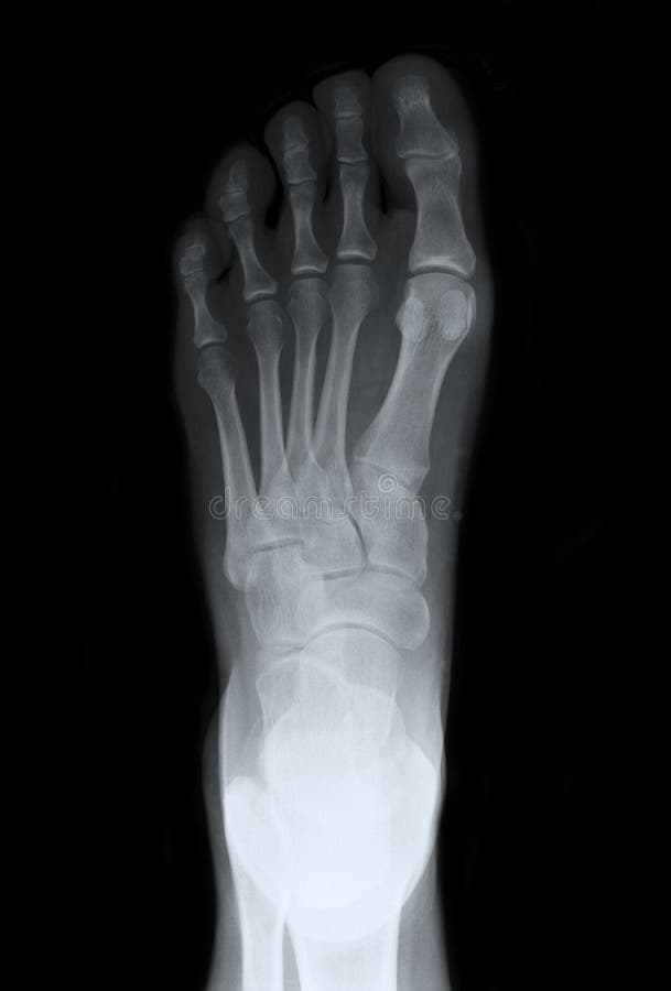
Left Foot Top Xray stock photo. Image of office, bones 23546186
The bases of the metatarsals and the tarsal bones are the most reliable rotation indicator on the DP view. If the foot is over rotated externally, the metatarsal bases will be heavily superimposed whilst the tuberosity of the navicular bone can be seen in profile. Over rotation internally will open up the metatarsal bases and the resultant.

Normal ankle xray netlaser
This is a basic article for medical students and other non-radiologists. A foot x-ray, also known as foot series or foot radiograph, is a set of two x-rays of the foot. It is performed to look for evidence of injury (or pathology) affecting the foot, often after trauma.
Standing lateral view Xray of the left foot. The os intermetatarseum... Download Scientific
This webpage presents the anatomical structures found on foot radiograph. Foot X-ray AP. Foot X-ray oblique. What Is Foot Radiograph? A foot radiograph or X-ray is a diagnostic imaging test that uses radiation to produce an image of the foot's bones and soft tissues (1).An X-ray image shows darker shades for the muscles and soft tissues, and the bones appear white in the film image (2).

Mortise and Lateral View Xray of Left Ankle. Mortise and lateral view... Download Scientific
Download scientific diagram | Left foot X-ray: (a) Anteroposterior view; (b) lateral view; (c) oblique view and (d) axial calcaneus view. Note the gross talar head irregularity with dense areas.
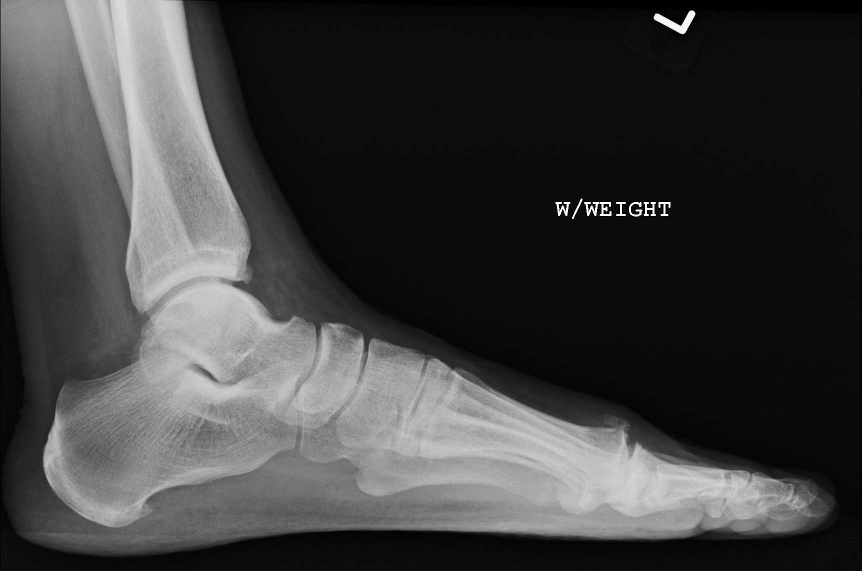
Figure 2
Lisfranc injury. The 'Lisfranc' ligament stabilises the mid-forefoot junction. Loss of alignment of the 2nd metatarsal base with the intermediate cuneiform indicates injury to this important ligament. Every post-traumatic foot X-ray must be checked for loss of alignment at the midfoot-forefoot junction (tarsometatarsal joints).

Normal foot xray ownnipod
Gender: Female. x-ray. Frontal. Oblique. Lateral. Normal right foot radiographs in a young adult female for reference.

Normal Left Ankle Xray
What to Expect During Your Foot X-Ray Procedure. Before the x-rays are taken you will be asked to take off your shoes and socks and roll up the legs of your pants. You will need to remove any jewelry or metal objects you may be wearing—for example, an ankle bracelet or toe ring. If you are pregnant, you must let the doctor know before.

Xray left foot metatarsal pain r/Radiology
Remember to check the whole film, though. Often, a foot x-ray is also requested for the investigation of osteomyelitis , arthritides , or bone lesion. This article relates mainly to traumatic injuries to the foot. A basic review should start with AP and lateral views (including the entire foot and ankle). With the exception of trauma, these.
:max_bytes(150000):strip_icc()/x-ray-image-of-bone-fracture-at-5th-metatarsal-left-foot-945203958-140a7bb8add94610838f0b3632543a5c.jpg)
Jones Fracture of the Foot Symptoms, Treatment, and Recovery
What is a Foot X-ray? A foot X-ray is a painless medical imaging technique that uses low levels of radiation to create detailed images of the bones and soft tissues in the feet. It's a non-invasive way to examine the internal structures of the feet, making it an essential tool for diagnosing various foot conditions.
Image
The foot series is comprised of a dorsoplantar (DP), medial oblique, and a lateral projection.The series is often utilized in emergency departments after trauma or sports related injuries 2,4.. See: approach to foot series. Indications. Foot radiographs are performed for a variety of indications including 1-4: . foot trauma
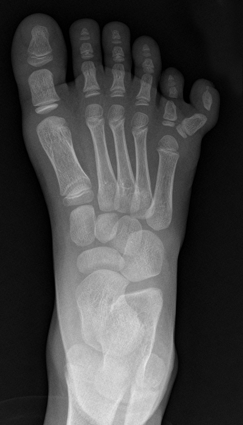
NORMAL FOOT 1
1. Check you have the right views. There are two views in foot x-rays DP (dorsal-plantar) and oblique. Both should ideally be done when weight-bearing if your patient can manage it. 2. Review the bones. Work round the bones one by one (including the metatarsals). Start proximally and work your way down, going medial lateral.… Read More »Foot x-rays

Normal ankle Image
This advanced CT is indicated for the evaluation of cortex and trabecular bone detail. This review discusses causes of chronic foot pain ( Table 1 2, 3; Figure 1), their clinical presentations.